Research
Cartilage
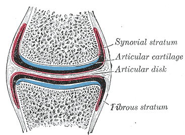
The Articular Cartilage at the end of the knee is a type of Hyaline cartilage. The cartilage consists of a matrix of collagen fibers, proteoglycan, elastin fibers, and cells called Chondrocytes. The extra-cellular matrix is maintained by the Chondrocytes, which rest in small pockets known as lacunae in groups of 2+. The collagen fibers function as the bulk connective proteins in the matrix, while proteoglycans work to attract water into the semi-permeable matrix to bring nutrients to the cells. Elastin functions to add elasticity to the matrix, and allow it to reform after undergoing high compressive loads.
Types
Cartilage consists of 3 main types that differ in the percentages of their proteins, which lead to different properties:
- Elastic
- Contains higher amounts of elastin to allow for more flexibility. Found mainly in the ears and epiglottis (flap connecting to the larynx).
- Hyaline
- Type found in the knee and commonly in the joints.
- Fibrocartilage
- Cartilage that contains more fibrous tissue and type 1 collagen (unique to Fibrocartilage). Found naturally in the body, as well as a potential form of scar tissue for damaged hyaline cartilage.
Bio-mechanical Properties
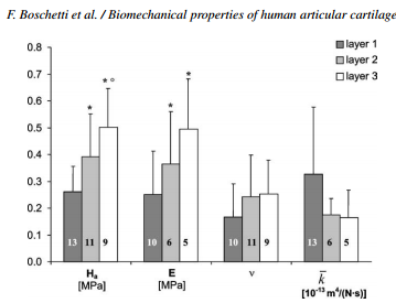
Cartilage is an elastic and semi-permeable material that has to undergo large compressive loads in order to support our joints. The measurable properties of cartilage vary based on depth, current stresses, and age, and generally are confined to the following 4 measures: 
- Young's Modulus (E)
- A measure of the stiffness of the material, in units of Stress/Strain. Young's Modulus is typically written in Mega-Pascals (MPa), and can be used to describe the amount of compression of the cartilage under a certain force
- Aggregate Modulus (Ha)
- The modulus that describes the stiffness of the material at a certain point, when there is no osmotic pressure (water isn't flowing in or out).
- Poisson's Ratio (v)
- The ratio of how much the cartilage compresses in the direction of the applied pressure, over how much the cartilage expands perpendicular to the direction of pressure.
- Hydraulic Permeability (k)
- The ease with which liquids can pass through the cartilage. Is dependent on the current pressure on the cartilage as well as individual factors.
External Links
Velcro
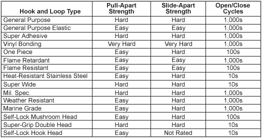
Industry Standards look at 3 different components of velcro strength: strength perpendicular to the plane of the velcro, or peel strength; strength horizontal to the velcro, the shear strength; and the number of times that the fasteners can be torn apart before losing strength. Typical hook and loop fasteners tend to have a lower peel and shear strength, but higher reusability. Other off brand fasteners that utilized bulb'd heads have been found to be significantly stronger, but have a very limited life after being reused.
External Links
McMaster-Carr's about velcro page
Industry Standard Test for the Shear Strength of Velcro
Video Demonstrating the Peel Strength Test
Paper that tested velcros using Shear, Lateral Shear, and Peel test methods
Bone Cement
Bone cements are materials that are used as an anchor for attaching prosthesis to bone. The cement is a form of Plexiglass, or PMMA (polymethyl methacrylate). The cement is stored in the form of a liquid and a powder that are vacuum-mixed before use. The resulting mixture will start as a liquid, and gradually become more solid as it sets, with the set time being variable based on the specific cement used.
Components
Powder
- Polymer (PMMA)
- Starter (Benzoyl Perdoxide initiator)
- Radiopacifiers (ZrO2/BaSO4)
- Antibiotics
Liquid
- Monomer (MMA)
- Initiator (N,N-dimethyl-p-toluidine)
- Inhibitor (Hydroquinone)
Procedure
- Clean Bone Bed
- Ensures maximum surface area in connection
- Mix Bone Cement
- Uses an Automatic Mixer or special vacuum mixing apparatus
- Apply Cement
- Must be done during set time allotted by specific cement
- Set prosthetic
- Pressurize Area
- Area must be pressurized until cement is strong enough to resist being pushed out by the blood pressure in the body
Providers
| Cement Brand | Manufacturer | Website |
|---|---|---|
| Allegiance | Cardinal Health, McGaw Park, IL | www.cardinal.com |
| BoneSource | Stryker Leibinger, Kalamazoo, MI | www.strykercorp.com |
| Cranioplastic | Depuy International, Ltd., Blackpool, England | www.codmanjnj.com |
| Orthocomp | Orthovita, Malvern, PA | www.orthovita.com |
| Osteobound | Zimmer, Warsaw, IN | www.zimmer.com |
| Placos | E. Merck, Darmstadt, Germany | www.merck.com |
| Palacos E-Flow | Essex Chemie AG, Lucerne, Switzerland | www.essex.ch |
| Simplex P | Stryker-Howmedica-Osteonics, Mahwah, NJ | www.strykercorp.com |
| Sucour | ArthoCar, Sunnyvale, CA | www.arthocare.com |
| Vertifix | WE Cook, Bloomington, IN | www.cookgroup.com |
External Links
Research on Properties of Different Cements
Attachment of Artificial Cartilage to Underlying Bone
Mechanical strength of acrylic bone cements impregnated with antibiotics
Bone Glue
Sandworm SuperGlue
http://www.moleclues.org/news/sandworm-superglue-mend-broken-bones
- By copying the shape and charge of the molecules of the sandworm's natural glue, scientists have made synthetic glue which will set in wet environments, like inside our body. It can be sucked into a syringe and used to repair small bone fractures. The glue can also carry drugs, so it could simultaneously be used to fix the break and deliver ant-inflammatory drugs to the injured area. Eventually, the aim is to develop the glue to be biodegradable, so it would be replaced by healthy bone tissue.
http://www.sciencedaily.com/releases/2008/11/081125085620.htm
* The synthetic sandcastle worm glue would not be used to repair large fractures such as major leg and arm bones, for which rods, pins and screws now are used. But Stewart envisions that it might be used for gluing together small bone fragments in fractured knees, wrists, elbows, ankles and other joints, and also the face and skull.
http://www.nytimes.com/2010/04/13/science/13adhesive.html?pagewanted=all&_r=0\
- The worm produces the glue in two parts, with different proteins and side groups in each. The two are made separately in a gland, and, like an epoxy, come together only as they are secreted. When they mix they form a compound that, even though water based, does not dissolve. The glue sets initially in about 30 seconds, probably triggered by the abrupt change in acidity — it is far more acidic than seawater, Dr. Stewart said. Over the next six hours, the adhesive hardens completely as cross-links form between the proteins. “It turns into this thing that has the consistency of shoe leather,” he said. “It’s still flexible but very tough.”
Cyanoacrylates "Superglue"
http://www.loctitehf.com/assets/tds/NEW-CA401-EN.pdf
- Technical Data sheet of a commonly used bone glue
http://www.aspirebiotech.com/medical_grade_cyanoacrylates.html
- Aspire Biotech,Inc. is a producer of medical grade cyanoacrylate.
http://www.jniosh.go.jp/en/indu_hel/pdf/42-2-14.pdf
- A scientific paper from 2004 detailing the toxicity of cyanoacrylates in dental use
http://www.sirchie.com/assets/msds/cna102.pdf
- MSDS on Cyanoacrylates
http://www.paseca.com/images/Cyanoacrylate-Guide.pdf
- Complete guide on different dispensing techniques and equipment necessary for application of the glue. But this isn't a guide focused mainly on medicine but the general application of this.

